>ABSTRACT
Background The large caliber of head hair in hair transplantation imparts a coarse hairline, whereas natural hairlines are typically softer. In hirsute individuals, transplantation of leg hair to the hairline may result in a superior aesthetic appearance.
Observation A total of 1025 leg hair follicles in 1 patient were grafted to an area 0.5 to 1.0 cm in front of and 0.5 to 1.0 cm internal to the vanguard hair of the original hairline and temporal recesses; the other patient received approximately 1000 leg hairs and 600 head hairs to advance and soften his hairline and to create a custom widow’s peak. Transplantation resulted in a fully grown and soft-looking hairline after 9 months in the first patient, with growth of 75% to 80% of the transplanted leg hair. The mean length of the transplanted leg hair was longer than the length of the original leg hair, with less curliness but similar hair width. Transplanted leg hair width was significantly finer compared with existing head hair width. After 4 years, results were sustained, minimizing concerns that subsequent hair loss might result from leg hair cycle variations. In the second patient, similar results were observed at 3 years.
Conclusion The use of leg hair in transplantation provides additional options in patients with hairlines that need to be refined.
Hairlines created by modern hair transplant techniques use single hair follicles derived from the safe donor area (SDA). Swinehart1(p868) aptly noted that “a normal hairline is not a line at all, but rather a zone in which hairs become finer and more sparse as they gradually give way to the hairless forehead skin. This diminution in diameter and density is most prominent in younger patients, along temporal hairlines, and in female patients.” In a transplanted hairline, which aims to mimic mild recession (Norwood class 2 or Norwood class 3), this observation becomes even more true. A receded hairline by nature assumes some miniaturization of the vanguard hair population.
The creation of a natural-looking hairline thus must include in the planning stages a deliberate attempt to add finer caliber hair in the very front of the hairline. Conventional hair transplantation, however, uses hair from the SDA that contains the thickest caliber hair in the entire scalp. In patients with especially thicker caliber hairs (eg, Asians) and in patients with darker hair color and a contrasting lighter skin color, the production of a natural, soft-looking hairline has remained a major challenge. The recognition of this problem has led to the grafting of bisected hair follicles into the hairline on the premise of thinner caliber hair growth. In the study performed by Swinehart1 using bisected hair follicles, wider application was limited by the poor yield (< 50%).
The advent of follicular unit extraction (FUE)2, which allows for in vivo extraction of single follicles without creating cosmetically significant scarring, has resulted in good results obtained in the frontal hairline. In addition, the technique increases the prospect of using single nonhead hair donor sources in transplantation3-6. The FUE technique harvests hair follicle grafts through the use of punch devices to incise a circular path around individual follicular units from the epidermis into the dermis. However, the wider variation in the caliber of hair in the different body areas (finer leg hair to coarser beard hair) has created the possibility of a new approach to softer hairlines in the field of hair transplantation, particularly when head donor hair is not an option. For example, leg hair in most individuals possesses a finer caliber compared with most scalp hair populations. The use of leg hair in transplantation has only been reported once, to my knowledge; Jones6 described the growth of leg hair in a hair transplant donor scar. The current report describes 2 patients with leg hair used to impart soft, natural-looking hairlines.
>REPORT OF CASES
PATIENT 1
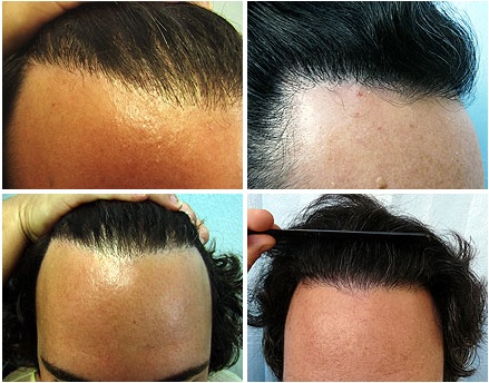
A 35-year-old white man with a history of multiple follicular unit strip surgery (FUSS), also called follicular unit transplantation (FUT), including implantation of single head follicles and micrografts to the hairline, presented with complaints of a harsh-looking hairline (Figure 1A). He was self-conscious of the problem and resorted to styling his hair forward to obscure the hairline. From his leg areas, 1025 leg hair follicles were extracted and grafted to an area covering 0.5 to 1 cm in front of and 0.5 to 1 cm internal to the original vanguard hair of the original hairline and temporal recesses (halfway in between the hairs in the existing hairline and the other half in front of the hairline). The hairline was fully grown and soft looking by 9 months, at which time the patient started combing his hair backward and sporting a pony tail, exposing his hairline comfortably. Approximately 75% to 80% of the transplanted leg hair grew. The result at 4 years revealed sustained results (Figure 1B). By 3 months the leg donor area had healed without an appearance of scars (Figure 2). In addition, hair length measurements for a comparable duration of the last hair cut also showed that the leg hair length on the hairline had a mean length of 4.0 cm, while leg hair on the leg had a mean length of 3.5 cm. In the same amount of time, transplanted head hair in the hairline had attained a mean length of 11 cm. Also, the leg hair on the hairline was less curly than leg hair on the leg.
Figure 1. Patient 1. A, Prior to surgery to refine the hairline. B, Four years after transplantation of leg hair, there is softening of the hairline.

Figure 2. Patient 1. A, Donor site (leg) immediately after hair extraction. B, Donor site (leg) 3 months later, completely healed and without scars.

>PATIENT 2
A 29-year-old white man had undergone FUSS/FUT and was unhappy about his hairline, which he felt was too harsh and straight. He had resorted to cropping his hair short to obscure the problem. He requested advancement and softening of his hairline, as well as the creation of a custom, accentuated widow’s peak. Approximately 1000 leg hairs and another 600 head hairs were used in combination (with more leg hair in the vanguard area than head hair to advance and soften) and create the custom widow’s peak. Approximately 75% to 80% of the transplanted leg hair grew. At 3 years, the patient’s result was sustained, allowing for a longer hair cut and the ability to comb the hair backward, exposing a softer hairline). The leg area extraction sites healed without an appearance of scars.
PROCEDURE
Both patients were hirsute with indications for use of nonhead hair for hair transplantation. In the case of donor hair extractions, leg and thigh donor areas were pretreated with minoxidil, 5%, once or twice daily for a variable period of 6 weeks to 6 months before surgery, and anagen hair was specifically used, preshaving the areas 7 to 10 days prior to surgery.
The procedure was performed under local anesthesia by subcutaneous injections of epinephrine (1:100 000) and lidocaine, 1%, and bupivacaine hydrochloride, 0.25%, in a 5:1 ratio for recipient areas, and a further dilution (5:1) with normal saline for donor areas. Tumescence was not performed in the donor areas. A rotary tool was used to mount modified hypodermic needles (19- and 20-gauge) that had tips to form a customized punchlike instrument. Individual hair follicles were excised using the sharp rotating needle tips to a depth exceeding the bulge area. Freed hair follicles were easily pulled out with occasional aid of blunt needle tip dissection.
Because wounds created by the customized needle tip widen with depth, as observed in these and numerous other cases, injury to follicles is diminished and wound closure accelerated. The wounds created tended to have inverted or straight edges favoring faster healing than in substantially everted wound edges. For recipient grafting, slits were created by means of blades custom sized to the dimensions of the extracted grafts. The time required for surgery was 5 to 6 hours for 1000 grafts.
COMMENT
These case reports demonstrate the successful transplantation of leg hairs to the hairline in 2 men who had undergone prior FUSS-FUT surgical procedures but who were dissatisfied with the unaesthetic results. In both instances, the source of dissatisfaction was in part from the use of thick caliber hair from the SDA of the head, which imparted a harsh hairline appearance. The primary advantage of using leg hair to rework hairlines is that it is relatively finer and thus can be used to fill in the vanguard regions of the hairline to create a much softer and more natural look.
In using body hair for transplantation to the head it is important to select actively growing anagen hair. In telogen hair, the lower one-third portions of the hair follicles have undergone apoptosis and are survived by a perifollicular sheath that forms a fibrous streamer comprised of fibroblasts, small blood vessels, and collagen.7 The scoring process in FUE and subsequent dissection and pulling of the follicle that it entails is too traumatic for a follicle whose lower one-third has degenerated. Moreover, at this stage of the hair cycle the follicle is more liable to be damaged (transected), leading to low yield. Because leg hair has a higher proportion of hair in the telogen phase compared with head hair (40%-85% vs <15%), I devised a protocol that maximizes the number of leg hairs in anagen that is more likely to better withstand the trauma of FUE. Early-stage anagen hair (I-IV) can also be mistaken for telogen hair owing to associated bulbs that have yet to become pigmented. Thus, one needs a method to indentify later-phase anagen hair, which is why preshaving the donor area is undertaken 3 to 7 days prior to extraction. Only hair that has grown above the skin is harvested because this would be actively growing hair and more likely to be in anagen (growth phase) of their cycle. This method of anagen hair selection borrows from the procedure of the phototrichogram, which was used in anagen hair studies first described by Saitoh et al8. These authors described anagen selection by counting growing hair 2 or more days after the area had been shaved to the skin8.
Scalp and leg hair growth cycles are also quite different. Whereas head hair is typically 85% to 90% in the anagen phase and this phase lasts several years on average, body hair has a much higher proportion of telogen hair (40%-85%), and the duration of anagen hair is much shorter and is measured in months9- 11. Preparation of donor sites with topical minoxidil shortens the telogen phase by induction of resting hair follicles into the anagen phase12. However, such use of minoxidil in preparation for transplants of body hair to the head has not been formally studied, and therefore this particular role needs further research. Because one patient was observed over a 4-year period and the other for 3 years with sustained results, the concern regarding shorter anagen hair cycles in leg hair, which might cause loss of transplanted hair, was eliminated. Based on these and other cases, it seems that sustained body hair on the body correlates with sustained growth of equivalent body hair on the scalp.
In both patients the yield from transplantation of leg hair was estimated at 75% to 80%, which is consistent with my findings in tests described in an earlier report using chest hair.3 A previous report in the literature indicated that a 4-fold increase in the length of chest hair was possible when it was transplanted to the scalp,5 but observations from these 2 patients suggest that the same phenomenon does not occur with leg hair. In this instance, the ultimate hair length was only slightly longer than the original leg hair. Folliscope images showed that the original mean leg hair width and transplanted mean (SD) leg hair width to the head were similar: 0.105 (0.022) mm (8 hairs in each set of measurements) vs 0.092 (0.015 mm) (6 hairs in each set of measurements), respectively (t test; P = .21). However, these hair calibers are in sharp contrast to the caliber of original head hair, which had a mean of 0.129 (0.009) mm (6 hairs in each set of measurements). The difference in hair width means between original head hair and leg hair transplanted to the scalp was significant (P < .001; t test). However, the curliness of the original leg hair in the first patient was much reduced. While this could be a nascent change in curliness, it is possible that the result simply reflects grooming practice—head hair is groomed, and leg hair is not. Although the maintenance of transplanted hair caliber is in accordance with the principle of donor dominance, first defined by Orentreich13 as the retention of donor hair characteristics at recipient sites, the observation that transplanted leg hair grows longer and straighter on the head may also be the result of some influence of the recipient site as demonstrated by several studies14- 16.
While published reports of the use of nonhead hair for transplantation to the scalp are still scant,3,5- 6 the present cases further emphasize the possibility of a new pool of donor supply for situations in which a patient has extensive baldness and insufficient head donor hair due to the extensiveness of the baldness, previous surgical procedures, or accidents, such as burns. Moreover, the variety of types of nonhead hair available enables a tailoring of the transplantation to match the desired characteristics of the target areas of the head. There are still some limitations or disadvantages to using FUE to transplant nonhead hair to the scalp. These include longer operating times, the need for a higher level of skill, variations in hair angulation, the likelihood that the quality of nonhead hair will not be as high as for head hair, and the need for sufficient body hair, which is typically found only in relatively hirsute individuals.
The most important application of this work is in selected hirsute individuals who have poor aesthetic results of harsh hairlines created previously using coarse terminal hair from the SDA of the head. This work also creates the possibility of creating more natural-looking, softer hairlines in individuals undergoing hair transplantation for the first time. In qualified individuals with larger caliber head hair, especially Asians and individuals with darker hair and sharply contrasting lighter skin color, this may be especially relevant. Because leg hair is finer and shorter at full length than scalp hair, this kind of hair may also be of use in the transplantation of eyebrows and eyelashes in which these nascent characteristics would fit more naturally. However, because great variation exists in eyebrow and eyelash caliber between individuals, this may not be a viable option in every instance.
>ARTICLE INFORMATION
Correspondence: Sanusi Umar, MD, FineTouch Dermatology Inc, 819 N Harbor Dr, Ste 400, Redondo Beach, CA 90277 (drumar@dermhairclinic.com).
Accepted for Publication: October 8, 2011.
Financial Disclosure: None reported.
This article was corrected for errors on May 8, 2012.
REFERENCES
1 Swinehart JM. “Cloned” hairlines: the use of bisected hair follicles to create finer hairlines. Dermatol Surg. 2001;27(10):868-872
PubMed
2 Rassman WR, Bernstein RM, McClellan R, Jones R, Worton E, Uyttendaele H. Follicular unit extraction: minimally invasive surgery for hair transplantation. Dermatol Surg. 2002;28(8):720-728
PubMed
3 Umar S. Hair transplantation in patients with inadequate head donor supply using nonhead hair: report of 3 cases. Ann Plast Surg. 2011;67(4):332-335
PubMed
4 Hirai T, Inoue N, Nagamoto K. Potential use of beards for single-follicle micrografts: convenient follicle- harvesting technique using an injection needle. Ann Plast Surg. 2001;47(1):37-40
PubMed
5 Woods R, Campbell AW. Chest hair micrografts display extended growth in scalp tissue: a case report. Br J Plast Surg. 2004;57(8):789-791PubMed
6 Jones R. Body hair transplant into wide donor scar. Dermatol Surg. 2008;34(6):857PubMed
7 Cotsarelis G, Millar SE. Towards a molecular understanding of hair loss and its treatment. Trends Mol Med. 2001;7(7):293-301
PubMed
8 Saitoh M, Uzuka M, Sakamoto M. Human hair cycle. J Invest Dermatol. 1970;54(1):65-81 PubMed
9 Ort RJ, Anderson RR. Optical hair removal. Semin Cutan Med Surg. 1999;18(2):149-158 PubMed
10 Straub PM. Replacing facial hair. Facial Plast Surg. 2008;24(4):446-452
PubMed
11 Randall VA. Androgens and hair growth. Dermatol Ther. 2008;21(5):314-328
PubMed
12 Yoo HG, Chang IY, Pyo HK, et al. The additive effects of minoxidil and retinol on human hair growth in vitro. Biol Pharm Bull. 2007;30(1):21-26
PubMed
13 Orentreich N. Autografts in alopecias and other selected dermatologic conditions. Ann N Y Acad Sci. 1959;83(3):463-479
PubMed
14 Hwang S, Kim JC, Ryu HS, et al. Does the recipient site influence the hair growth characteristics in hair transplantation? Dermatol Surg. 2002;28(9):795-799PubMed
15 Lee SH, Kim DW, Jun JB, Lee SJ, Kim JC, Kim NH. The changes in hair growth pattern after autologous hair transplantation. Dermatol Surg. 1999;25(8):605-609PubMed
16 Hwang ST, Kim HY, Lee SJ, Lee WJ, Kim W, Kim JC. Recipient-site influence in hair transplantation: a confirmative study. Dermatol Surg. 2009;35(6):1011-1014
For more hair transplant related publications by Dr Umar, click here